Radiology Videos
Items 1 to 10 of 19 - Click image to start video
 Dr. Mary Mahoney discusses ultrasound imaging of the breast.
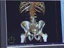
A CT scan showing the placement of a stent in the abdomen. The stent keeps the aneurysm from breaking.

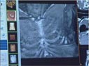
CT angiography images of the brain and arteries to the brain are captured and then reviewed by a radiologist. Angiography can be combined with CT to produce pictures of major blood vessels throughout the body. Angiography uses a contrast material to produce detailed images of both blood vessels and tissues. For more information see CT Angiography (CTA).
CT scans of the lungs and lung vessels are captured and then reviewed by a radiologist in order to identify a blood clot. CT is commonly used to assess for pulmonary embolism (a blood clot in the lung vessels).
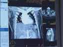
 3-D manipulation software is applied for different views and to remove visual layers of skin and bone as seen in these CT images of the chest.
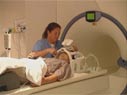
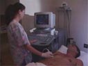 A radiologic technologist performing an internal organ Doppler ultrasound procedure on a patient. The procedure can be used to detect abnormal blood vessels and flow. For liver transplants it can assess blood flow through the portal vein.
 A radiologic technologist preparing a patient for a chest x-ray. A chest x-ray produces images of the heart, lungs, airways, blood vessels and the bones of the spine and chest.
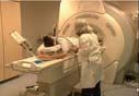 A radiologic technologist preparing a patient for a magnetic resonance (MR) imaging breast examination, commonly called breast MRI, and the technologist in the control room monitoring images during the procedure.
 A radiologic technologist preparing a patient with head gear for an fMRI exam and moving him through the magnetic resonance (MR) scanner as another technologist monitors the images in the control room.
 A radiologic technologist preparing a pediatric patient for a chest x-ray. A chest x-ray produces images of the heart, lungs, airways, blood vessels and the bones of the spine and chest.
