Images & Videos: CAT Scan (CT) - Chest
Items 1 to 10 of 36 - Click images to view larger
 CT scans of the aorta and coronary, or heart, blood vessels are captured and then reviewed by a radiologist.
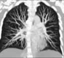
 3D Reconstruction Images of CT of lung.
CT angiogram. Frontal or coronal view of chest-3D slab image showing pulmonary vessels. For more information see CT Angiography (CTA).
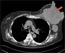
Two CAT (CT) scan 'slices' of the chest viewed in different settings showing a large breast tumor (red arrow).

Computed tomography (CT) scan in patient with chronic bronchitis showing thickening of the bronchial walls (red arrows) and mucous within the bronchi (yellow arrows).
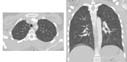
 3-D manipulation software is applied for different views and to remove visual layers of skin and bone as seen in these CT images of the chest.
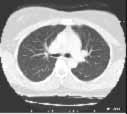
CT of the lungs, window level set to demonstrate the vessels and air ways - not intended to demonstrate the heart, spine muscles etc. This is used to look for things like pneumonia or lung cancer. For more information see Pneumonia.
Normal chest CT
