Images & Videos: Lower GI Tract X-ray
Items 1 to 9 of 9 - Click images to view larger
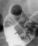
This image shows the right side of the large intestine. Air (dark) distends the bowel and barium (white) coats the inner lining.
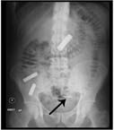
X-ray of the belly, frontal view: bright objects represent foreign bodies (batteries) which the patient swallowed. An additional dense line (arrow) in the lower right part of the image represents another metallic foreign body.
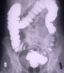
Normal air contrast barium enema
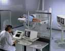
Radiography equipment
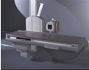
Radiography equipment
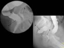
Sagittal images from a barium enema show the bright contrast instilled in the rectum (A) which also fills the vagina (B) through a fistula.
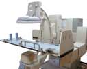
X-ray equipment is mounted on a C-shaped gantry with the x-ray tube itself beneath the table on which the patient lies. Above the patient is an image intensifier that receives the x-ray signals, amplifies them, and sends them to a TV monitor.
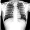
Chest x-ray demonstrating miliary Tuberculosis which is a diffuse form of Tuberculosis. Its name comes from a distinctive pattern seen on a chest X-ray of many tiny (white) spots distributed throughout the lung fields with the appearance similar to millet seeds, thus the term "miliary" tuberculosis. When the mycobacterium which causes the disease gets into the blood stream it can settle in many areas of the lung (and other organs) causing this miliary appearance.
