Images & Videos: Children's (Pediatric) Ultrasound - Abdomen
Items 1 to 10 of 10 - Click images to view larger

Ultrasound equipment
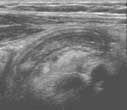
Ultrasound of appendicitis in a 5-year-old girl. The long banana-shaped structure in the center of the ultrasound image is an inflamed appendix indicating appendicitis.
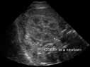
Sonogram - short axis or transverse view of the right kidney in a newborn.
 Your Radiologist Explains Pediatric Sedation and Anesthesia
Ultrasound equipment
 Your Radiologist Explains Imaging Appendicitis in Children
 Your Radiologist Explains Pediatric Radiology
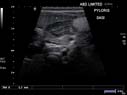
Ultrasound of a baby's belly shows abnormal thickening of the wall at the exit of the stomach, which is also called pylorus. The wall thickening blocks content of the stomach from moving on into the small bowel and therefore can cause vomiting (diagnosis of hypertrophic pyloric stenosis).

Pediatric radiologist scanning a boy's abdomen using ultrasound.
