Images & Videos: CAT Scan (CT) - Body
Items 1 to 10 of 32 - Click images to view larger
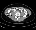
Appendicitis: A CT cross-section through the patient's lower abdomen has also cut through the swollen, inflamed appendix in two places (arrows). Inflammation has spread to the nearby intra-abdominal fat (arrowhead). For more information see Appendicitis.
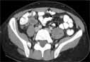
Appendicitis: The appendix (A) is distended and inflamed. In this patient the appendix has not yet ruptured. For more information see Appendicitis.

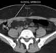
CT scan of a normal appendix in the right lower abdomen. The appendix normally connects with the right colon and contains air (this appears black on the scan). Air in the appendix excludes appendicitis since this means that the appendix is not obstructed or inflamed. For more information see Appendicitis.
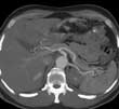
Reformatted image of the mid-abdomen creating a 'slab' showing the liver and intestines. The abdominal aorta and branches going to the liver, spleen and stomach are visible. This view is looking up toward the head of the patient.
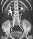
 Coronal Reconstruction of the Abdomen and Pelvis.
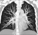
CT angiogram. Frontal or coronal view of chest-3D slab image showing pulmonary vessels. For more information see CT Angiography (CTA).
 Your Radiologist Explains Body CT/CAT Scan
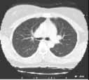
CT of the lungs, window level set to demonstrate the vessels and air ways - not intended to demonstrate the heart, spine muscles etc. This is used to look for things like pneumonia or lung cancer. For more information see Pneumonia.
