Images & Videos: Myelography
Items 1 to 8 of 8 - Click images to view larger

Myelography of lumbar spine - injection of contrast into spine to study disc disease.
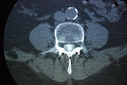
Contrast-enhanced computed tomography (CT) scan of a dural tube showing small black dots (arrows) that represent nerve roots, called the cauda equine.
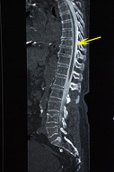
Contrast-enhanced computed tomography (CT) scan of the dural tube (arrow) in a patient’s spine.
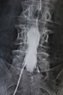
Image of a myelographic needle containing contrast inserted in the dural tube of a patient’s spine.
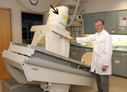
Image of a fluoroscopic table slightly tilted with an image intensifier (the 'x-ray eye') denoted by the arrow.
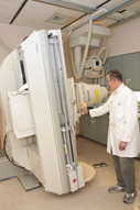
Image of a fluoroscopic table tilted in an upright position.

Radiography equipment
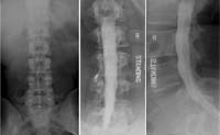
Preliminary view (1) of lumbar spine. Front (2) and side (3) views during a myelogram with iodine in the spinal canal.
