Images & Videos: CAT Scan (CT) - Head
Items 1 to 10 of 29 - Click images to view larger
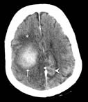
Brain metastasis: A cross-section through a patient's brain after administration of intravenous contrast shows the contrast accumulating in the periphery of a cancerous deposit that has traveled to the brain, giving a characteristic ring-enhancing appearance.
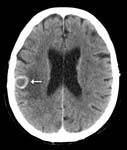
A CT cross-section through a patient's brain shows an intraparenchymal hemorrhage (arrow), meaning blood within the substance of the brain. Also shown is a subarachnoid hemorrhage (arrowhead), or blood surrounding the surface of the brain; and edema (asterisk), or swelling of the brain.


 2 brain aneurysms on a rotating 3D reconstruction.
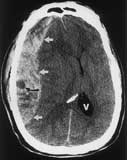
A computed tomography (CT) cross section of a patients head showing a large blood clot (arrows) located outside the brain, and called an acute subdural hematoma.
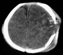
A computed tomography (CT) cross section of a pediatric patient's head showing an enlarged fracture in the skull denoted by the gap in the white circle. The speckled appearance inside the gap indicates bleeding. Outside the gap, the scalp tissues are swollen.
