Images & Videos: RFA (for Liver Tumors)
Items 1 to 6 of 6 - Click images to view larger
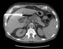
CT-guided radiofrequency ablation of a liver tumor: A cluster electrode is positioned in the patient's hepatocellular carcinoma. For more information see Radiofrequency Ablation of Liver Tumors.
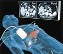
Computer-generated image depicts the elements of ultrasound-guided percutaneous ablation of liver tumors as well as representative pre- and post-ablation CT scans.
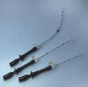
Radiofrequency ablation needle devices that contain multiple curved, retractable electrodes. The electrodes are kept inside the needle until its tip is positioned within a tumor.
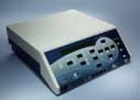
The radiofrequency generator. Connected by insulated wires to the needle electrode, the generator produces alternating electrical current in the range of radiofrequency waves.

Closeup of retractable electrodes used in radiofrequency ablation. A generator, shown in the background, provides electrical current for the electrodes.
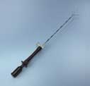
Radiofrequency ablation needle device that contains multiple curved, retractable electrodes. The electrodes are kept inside the needle until its tip is positioned within a tumor.
