Images & Videos: Scrotal Ultrasound
Items 1 to 6 of 6 - Click images to view larger
Ultrasound equipment
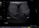
A scrotal ultrasound image showing normal testes (medium gray oval structures in the image, showing only part of each testicle).
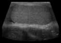
Scrotal sonogram showing side view (longitudinal) of a testicle. Patient's head to left and feet to right out of the field of view.
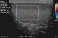
Color flow sonogram of one testicle, showing normal blood flow. Looking from the side. Patient's head is to the left and his feet are to the right.
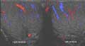
Sonogram of both testicles, with color showing blood flow. Looking up from the feet of a patient toward his head.
