Images & Videos: Computed Tomography (CT or CAT Scan)
Items 1 to 10 of 223 - Click images to view larger
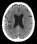
Brain metastasis: A cross-section through a patient's brain after administration of intravenous contrast shows the contrast accumulating in the periphery of a cancerous deposit that has traveled to the brain, giving a characteristic ring-enhancing appearance.
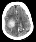
A CT cross-section through a patient's brain shows an intraparenchymal hemorrhage (arrow), meaning blood within the substance of the brain. Also shown is a subarachnoid hemorrhage (arrowhead), or blood surrounding the surface of the brain; and edema (asterisk), or swelling of the brain.
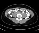
Appendicitis: A CT cross-section through the patient's lower abdomen has also cut through the swollen, inflamed appendix in two places (arrows). Inflammation has spread to the nearby intra-abdominal fat (arrowhead). For more information see Appendicitis.

A child being prepared for a CT scan. For more information see Children's (Pediatric) CT.
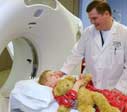
A child being prepared for a CT scan. For more information see Children's (Pediatric) CT.
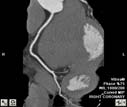
2D reconstruction image of a normal right coronary artery.
 CT scans of the aorta and coronary, or heart, blood vessels are captured and then reviewed by a radiologist.
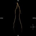
Normal three-dimensional CT angiogram of the lower extremities. For more information see CT Angiography (CTA).
 3D Reconstruction Images of CT of lung.
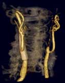
3D reconstruction image of the carotid arteries (CTA) showing a stent in right carotid and a 99% stenosis (blockage).
