Images & Videos: Breast MRI
Items 1 to 10 of 13 - Click images to view larger
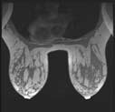
High-resolution MR image of both breasts provides anatomic detail.
An MR image of two breasts demonstrating vascular supply.
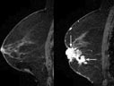
Contrast (a gadolinium-based agent) is administered during MRI of the breast to show areas suspected of being cancer which demonstrate contrast enhancement (appear brighter compared to rest of the breast tissue) as seen in the image (arrows).
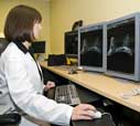
 A radiologic technologist preparing a patient for a magnetic resonance (MR) imaging breast examination, commonly called breast MRI, and the technologist in the control room monitoring images during the procedure.
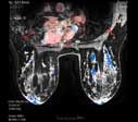
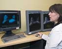
 A radiologist reviewing multiple images of the breast MRI and identifying cancer in the left and right breasts. The radiologist then adds the color overlay to show the contrast material being absorbed by the cancers.
 Your Radiologist Explains MRI of the Breast (Breast MRI)
 Dr. Mary Mahoney discusses breast MRI.

MRI scan of the breast
Breast MR procedure
Breast MR procedure
|
next 3 items|
