Images & Videos: Abdominal and Pelvic CT
Items 1 to 10 of 32 - Click images to view larger


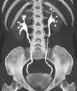
A CT scan showing the placement of a stent in the abdomen. The stent keeps the aneurysm from breaking.
Appendicitis: The appendix (A) is distended and inflamed. In this patient the appendix has not yet ruptured. For more information see Appendicitis.
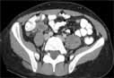
 3D modelling of CT (computed tomography) images of the abdomen in a cancer patient.
 Axial CT of Abdomen and Pelvis Normal study.

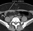
CT scan of a normal appendix in the right lower abdomen. The appendix normally connects with the right colon and contains air (this appears black on the scan). Air in the appendix excludes appendicitis since this means that the appendix is not obstructed or inflamed. For more information see Appendicitis.
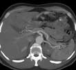
Reformatted image of the mid-abdomen creating a 'slab' showing the liver and intestines. The abdominal aorta and branches going to the liver, spleen and stomach are visible. This view is looking up toward the head of the patient.
