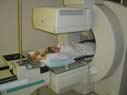
Images & Videos: Nuclear Medicine
Items 1 to 10 of 46 - Click images to view larger

Gastrointestinal bleeding: The dark spots moving on the right side of the image indicate that this patient is actively bleeding into the right colon. This is important information for the doctor to know in determining the appropriate treatment.

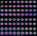
A PET/CT scan volumetric image demonstrating diffuse FDG accumulation in the bone marrow due to recent chemotherapy in a non-hodgkin lymphoma patient.
 Your Radiologist Explains Breast Lymphoscintigraphy
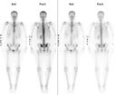
Colorectal carcinoma in a patient with rising carcinoembryonic antigen tumor marker levels.
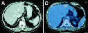
A = Computed tomography (CT) scan with intravenous contrast not showing metastatic disease.
B = Horizontal positron emission tomography (PET) scan showing focal abnormal radiopharmaceutical uptake in right lobe of liver.
C = Biopsy showing liver metastasis.