Images & Videos: Musculoskeletal MR
Items 1 to 7 of 7 - Click images to view larger
 Your Radiologist Explains MR Arthrography of the Hip
 Your Radiologist Explains MR Arthrography of the Shoulder
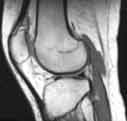
MR of the knee - side (lateral) view, showing distal or lowest part of femur, the patella (knee cap) and proximal (upper) tibia. The lateral meniscus is seen as a dark bow-tie like structure. The patellar tendon is also clearly seen at the front of the knee connecting the patella with the tibia.
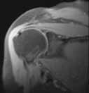
MR of the right shoulder looking at rotator cuff and head of humerus as well as glenoid portion of scapula.
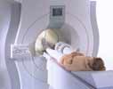
Magnetic Resonance Imaging (MRI) procedure
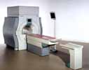
Magnetic Resonance Imaging (MRI) equipment
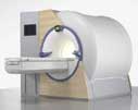
Magnetic Resonance Imaging (MRI) equipment
