Images & Videos: Ultrasound (Sonography)
Items 1 to 10 of 116 - Click images to view larger


Ultrasound equipment
 Your Radiologist Explains Abdominal Ultrasound (Sonography)
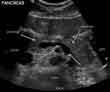
Normal pancreas seen on sonogram. Looking up from abdomen toward the head of the patient. The liver is in front of the pancreas. A vein draining the spleen is behind the pancreas.
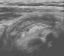
Ultrasound of appendicitis in a 5-year-old girl. The long banana-shaped structure in the center of the ultrasound image is an inflamed appendix indicating appendicitis.
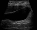
Ultrasound of the gallbladder. The wall or lining of the gallbladder is white, the bile within the gallbladder is mostly made of water and it appears as a clear black space within the walls of the gallbladder.
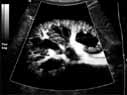
Power Doppler ultrasound of the kidney. This image shows the tiny blood vessels in the kidney like the branches of a tree.
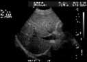
Ultrasound of the liver. This image demonstrates the liver tissue. The darker linear areas in the liver are veins bringing blood and nutrients to the liver and others are draining blood from the liver and returning it to the heart.
