Images & Videos: Upper GI Tract X-ray
Items 1 to 10 of 16 - Click images to view larger
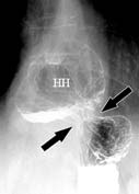
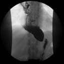
Upper GI x-ray image of a patient's diaphragm (arrows) showing a large hiatal hernia with high-volume reflux.
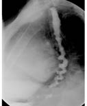
Lateral image from an esophagram demonstrates a "corkscrew" esophagus, consistent with diffuse esophageal spasm that can be seen in patients with a severe motility disorder of the esophagus.
Esophageal cancer seen on a barium swallow or upper GI exam (arrow).
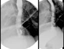
Spot radiograph (x-ray of a localized region) showing a normal gastroesophageal junction (straight arrow) and the diaphragmatic hiatus (curved arrow).
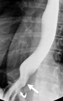
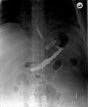
Dilated esophagus secondary to tight lower esophageal sphincter (achlasia)
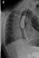
Image depicting esophageal narrowing due to cancer.
An x-ray image of the upper abdomen following oral contrast demonstrates a Lap-Band (a device that limits food intake) in good position.
Radiography equipment
|
next 6 items|
