Images & Videos: Intravenous Pyelogram (IVP)
Items 1 to 8 of 8 - Click images to view larger
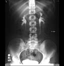
Contrast is seen in the kidneys, ureters, and bladder.
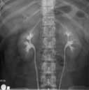
An x-ray image of the upper abdomen 10 minutes after the injection of contrast material shows normal kidneys, collecting systems and upper ureters.
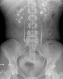
An x-ray image of the whole abdomen taken 15 minutes after injection of contrast material shows further excretion into the lower ureters and bladder.
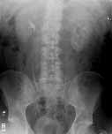
IVP at 5 minutes
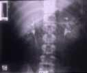
IVP, normal kidneys
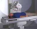
Radiography procedure
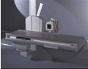
Radiography equipment
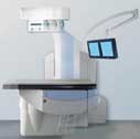
Suspended above the large, flat table is an apparatus containing the x-ray tube. The apparatus moves on a jointed 'arm' so that it can be properly positioned. Above the table to the right are video screens.
