Images & Videos: Bone X-ray
Items 1 to 10 of 43 - Click images to view larger
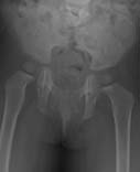
Plain x-ray image of a pediatric patient's pelvis. The image shows sacral agenesis (rare failure of sacral bone development).
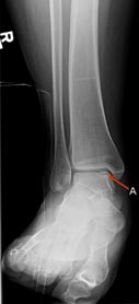
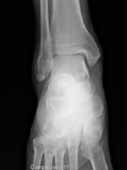
An x-ray image showing an osteochondral defect in the dome of the talus (a bone in the foot).
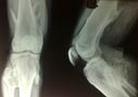
An x-ray image showing multiple hereditary exostoses.
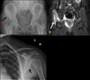
In this series of images, the letter A points to a lytic lesion in the right iliac (hip) bone. The letter B is an MRI scan showing the bone marrow edema (swelling) of the involved iliac bone. This was proved to be osteomyelitis (inflammation of the bone marrow and adjacent bone). The letter C points to a similar lesion that appeared in the left scapula (shoulder blade) a few months later. This is a Chronic Recurrent Multifocal Osteomyelitis (CRMO).
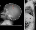
An x-ray image -- letter A shows a beveled lytic lesion seen in the skull. Letter B is vertebra plana. This is a case of Eosinophilic granuloma.
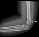
An x-ray image showing a posterior (rear) displacement of the posterior fat pad. This is an elbow joint effusion (fluid leak).
Ankle x-ray (front view)
 Your Radiologist Explains Bone X-ray
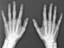
X-ray showing frontal view of both hands.
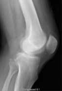
Knee x-ray (side view)
