Images & Videos: X-ray (Radiography)
Items 11 to 20 of 173 - Click images to view larger
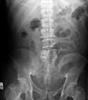
This image shows mild dilation of the small bowel gas.
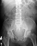
Normal abdomen.
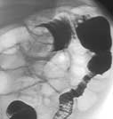
This image shows the right side of the large intestine. Air (dark) distends the bowel and barium (white) coats the inner lining.
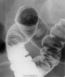
Liquid enema x-ray showing an intussusception in a pediatric patient.
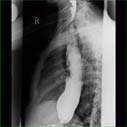
Side view from an esophagram shows an enlarged esophagus filled with contrast material with narrowing close to its connection to the stomach which is a common appearance in a condition called "achalasia." The esophagus of patients with "achalasia" does not relax normally because of an abnormality of the nerves in the wall of the esophagus and therefore it appears narrow.
 Your Radiologist Explains Airport Scanner Safety
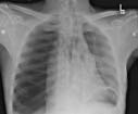
Frontal radiograph of the chest demonstrating a right tension pneumothorax.
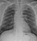
X-ray of a patient's chest showing a right-side aortic arch.
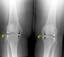
Knee x-ray of patient with osteoarthritis showing inner joint space narrowing (black arrows) due to cartilage loss and degenerative spur formation (curved arrows). The white arrows show normal space between the bones.

Musculoskeletal radiologist using fluoroscopic images to plan an ankle arthrogram.
