Images & Videos: Ultrasound (Sonography)
Items 11 to 20 of 116 - Click images to view larger
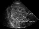
Sonogram - short axis or transverse view of the right kidney in a newborn.
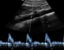
Ultrasound, abdominal aorta with Doppler wave form— side view: patient's head to left.
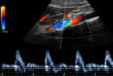
Ultrasound of abdominal aorta. Color flow and spectral doppler.
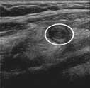
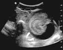
Ultrasound image of a swollen appendix in cross section shown as a rounded doughnut shape depicted in the circle.
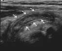
Ultrasound image of a swollen appendix shown lengthwise (arrows).
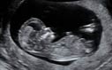
Ultrasound image of a fetus.
 Dr. Mary Mahoney discusses ultrasound imaging of the breast.

Dr. Mary Mahoney discusses biopsies.
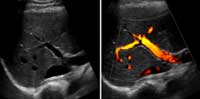
Ultrasound showing blood flowing from intestines into liver. Image on the left: routine. On the right with power Doppler.
Ultrasound image of a pediatric patient with anechoic (free of echoes) fluid between the two bowel walls of an intussusception.
