Images & Videos: Nuclear Medicine
Items 11 to 20 of 46 - Click images to view larger
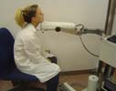
Photograph of a typical probe counter used for thyroid uptake exams. The patient sits with the camera directed at the neck for five minutes, and then the leg for five minutes.
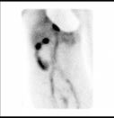
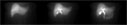
Positive GI bleeding scan with active bleeding site seen in the right colon.
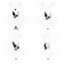
This is a thyroid scan that shows multiple irregular rounded bright areas within the thyroid. This represents a 'multi-nodular' goiter.
 Your Radiologist Explains PET Scans
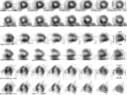
Normal Nuclear Medicine Myocardial (heart) Perfusion Scan. This scan is performed on a camera that rotates around the patient and produces 'slices' of the heart in several different views. The nuclear radiologist looks for areas in the heart muscle that are not receiving enough blood flow. These areas can often be treated to restore blood flow.
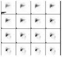
Normal hepatobiliary imaging.
Hepatobiliary imaging showing normal emptying of the gallbladder.
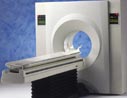
Positron Emission Tomography (PET) equipment
Normal Hepatobiliary ("HIDA") Scan. The liver is first seen alone with the bile duct and gallbadder noted on the later images.
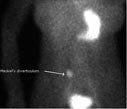
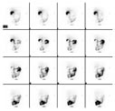
The bright spot in the image (indicated by an arrow) corresponds to a Meckel's diverticulum. The normal stomach is seen in the upper right and the normal urinary bladder is seen at the very bottom.
