Images & Videos: Magnetic Resonance Imaging (MRI)
Items 11 to 20 of 103 - Click images to view larger

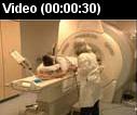
 A radiologic technologist preparing a patient with head gear for an fMRI exam and moving him through the magnetic resonance (MR) scanner as another technologist monitors the images in the control room.
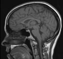
 Your Radiologist Explains MRI Scan of the Brain
 Your Radiologist Explains MR Angiography of the Brain and Neck
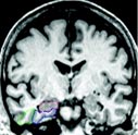
Figures A and B highlights three regions of the brain, including the:
-Hippocampus (outlined in red) that is responsible for forming memory, storing and organizing.
-Entorhinal cortex (outlined in blue) that is responsible for memory and navigation.
-Perirhinal cortex (outlined in green) that is responsible for object perception, memory and associations.
Figure A shows no atrophy. Figure B shows severe atrophy, with the exception of the right perirhinal cortex which has moderate atrophy.
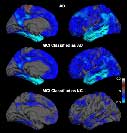
Image shows the average differences in brain thickness for subjects with Alzheimer’s disease (AD) and mild cognitive impairment (MCI). The blue areas indicate regions of thinning from the disease.
-Top: Healthy control (HC) subjects versus subjects with AD.
-Middle: HC subjects versus subjects with MCI who had AD imaging phenotype (an observable trait).
-Bottom: HC subjects versus subjects with MCI who had HC imaging phenotype.
 A radiologic technologist preparing a patient for a magnetic resonance (MR) imaging breast examination, commonly called breast MRI, and the technologist in the control room monitoring images during the procedure.
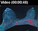 A radiologist reviewing multiple images of the breast MRI and identifying cancer in the left and right breasts. The radiologist then adds the color overlay to show the contrast material being absorbed by the cancers.
 Your Radiologist Explains MRI of the Breast (Breast MRI)
