Images & Videos: Pelvic Ultrasound
Items 11 to 16 of 16 - Click images to view larger
Uterus made with transvaginal probe - side view

Three-dimensional (3-D) ultrasound image of a patient's pelvis showing the endometrium.

Ultrasound image of a patient's pelvis showing the ovary.
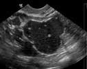
Ultrasound image showing ovarian teratoma.
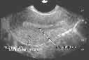
Intrauterine devices (arrow) are usually a safe means of contraception, but uncommonly they may push through the uterine wall (U), causing pain or other problems.
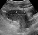
U = Uterine wall. The uterus is mostly dark on ultrasound, as shown on this sagittal (from the side) view.
U = Uterine wall. The uterus is mostly dark on ultrasound, as shown on this sagittal (from the side) view.
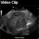 Real-time ultrasound scan through a uterus in a young woman.
