Images & Videos: Prostate MRI
Items 11 to 15 of 15 - Click images to view larger
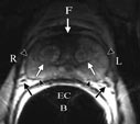
Endorectal MRI of the anatomy of the prostate using a clinical MR scanner. The anterior fibromuscular stroma (arrow) consists of nonglandular tissue and appears dark. The muscular stroma layer in the posterior prostate base is denoted by arrowheads.
F = Front
R = Right
L = Left
B = Back
EC = Endorectal Coil
F = Front
R = Right
L = Left
B = Back
EC = Endorectal Coil
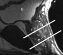
MRI image through the center of the male pelvis seen from the side. Bladder (B) is seen in the upper part of the image.
P = Prostate
U = Urethra
P = Prostate
U = Urethra
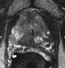
MRI scan in patient with prostate cancer. The cancer, marked with an arrow and the letter "C", appears as a rounded dark area.
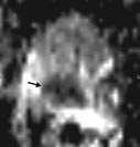
Diffusion weighted MR image shows how cancer (arrow), which appears as a dark area, inhibits the movement of water molecules in the area of the cancer.
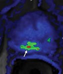
Computer generated MR image shows a perfusion abnormality in the area affected by cancer. The green and red areas (arrow) indicate that there is rapid uptake and washout of the contrast material which is seen in prostate cancer.
