Images & Videos: Ultrasound - General
Items 11 to 16 of 16 - Click images to view larger
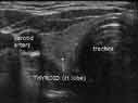
Sonogram of thyroid, right lobe, looking up from chin to the top of the head
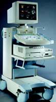
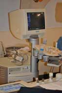
Ultrasound equipment
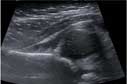
5 month old female: hip evaluated for congenital dysplasia
Ultrasound showing a dislocated hip--head of femur out of alignment with hip socket.
Ultrasound showing a dislocated hip--head of femur out of alignment with hip socket.
5 month old female: hip evaluated for congenital dysplasia
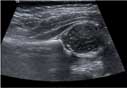
Ultrasound of a normal hip.
a. gluteus muscle
b. ilium
c. acetabulum
d. head of femur
Ultrasound of a normal hip.
a. gluteus muscle
b. ilium
c. acetabulum
d. head of femur

Ultrasound equipment
An ultrasound machine.
