Images & Videos: CAT Scan (CT) - Chest
Items 11 to 20 of 36 - Click images to view larger

Drs. Philip Alderson, Elliot Fishman and Robert Novelline discuss chest CT.
 Your Radiologist Explains CT of the Chest
 Your Radiologist Explains CT (CAT Scan) for Pulmonary Embolism
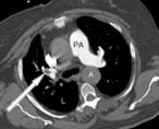
A pulmonary embolus (blood clot in the lung) within a pulmonary artery. The artery is white (because of high-density intravenous contrast), and the PE (arrow) is a dark glob within the artery.
PA = main Pulmonary Artery
L = Lungs (appear dark compared to the rest of the body because the air in lungs allows the xrays to pass through easily)
A = Aorta (the major artery leaving the heart)
PA = main Pulmonary Artery
L = Lungs (appear dark compared to the rest of the body because the air in lungs allows the xrays to pass through easily)
A = Aorta (the major artery leaving the heart)
 Patients undergoing a computed tomography (CT) procedure, sometimes called CAT scan, and a newer 64-slice CT scan. The 64-slice CT technology allows the radiologist a greater level of manipulation of 3-D clinical images to provide more complete anatomical views. These visuals and the clinical images can be used to report on CT scans of the body, chest, abdomen, vascular system and head.

CT image showing normal blood vessels within the lung.

Computed tomography (CT) scan showing large black areas within the upper lungs representing a severe case of emphysema.

Computed tomography (CT) scan showing black areas at the upper most part of the lungs representing emphysema.
