Images & Videos: Bone X-ray
Items 11 to 20 of 43 - Click images to view larger
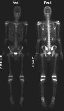
Bone Scan - Normal. Two pictures (one from the front and one from the back) of a normal bone scan from a 12-year-old boy.
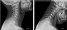
Lateral x-ray views of the cervical spine in extension (bending backward) and flexion (bending forward) demonstrating a normal spine alignment.
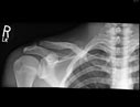
X-ray of the right shoulder area demonstrating a fracture of the clavicular bone.

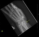
Plain x-ray image of a patient's left wrist showing a fracture of the scaphoid bone.
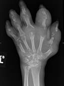
Plain film radiographic (x-ray) image (posterior-anterior view) of the left hand, demonstrating severe bony destruction and soft tissue swelling of the hand in a patient with longstanding gout.
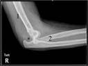
Side view of the elbow joint. The end of the upper arm (1) and the lower arm (2) are shown. The elbow joint is dislocated. The upper arm bone would normally be in line with the so-called olecranon process (*) of the lower arm bone.
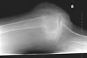
Plain x-ray image of a patient's knee showing fatty marrow in the joint space.
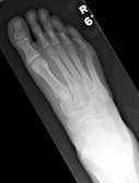
Plain film radiographic (x-ray) image (anterior-posterior view) of a normal right foot.
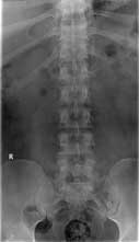
Normal frontal appearance of the lumbosacral spine
Frontal (A) and Lateral (B) radiographs of the left forearm and wrist demonstrate an intra-articular fracture of the radial styloid process (yellow arrow) with dorsal displacement of the fracture fragment and the carpal bones (red arrows). This fracture can be referred to as a 'combined Hutchinson (chauffeur) and dorsal Barton fracture' (a complex comminuted intra-articular fracture of the distal radius).
