Images & Videos: Magnetic Resonance Imaging (MRI) - Body
Items 11 to 20 of 24 - Click images to view larger
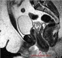
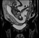
An MR image of an inverted, prolapsed uterus.
 Your Radiologist Explains MRI of the Body
Gallstone causing bile duct obstruction.
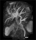
Magnetic resonance pancreatography showing a gallstone (arrow) obstructing the common bile duct.
Magnetic resonance pancreatography showing a gallstone (arrow) obstructing the common bile duct.
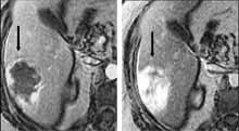
Benign hemangioma of the liver.
Gadolinium-enhanced MR image of the liver shows a enhancing lesion (arrow) that enhanced peripherally early (left) and fills in with contrast over time.
Gadolinium-enhanced MR image of the liver shows a enhancing lesion (arrow) that enhanced peripherally early (left) and fills in with contrast over time.
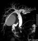
MRCP image: shows markedly dilated bile ducts and gallbladder from obstruction.
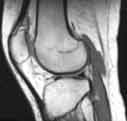
MR of the knee - side (lateral) view, showing distal or lowest part of femur, the patella (knee cap) and proximal (upper) tibia. The lateral meniscus is seen as a dark bow-tie like structure. The patellar tendon is also clearly seen at the front of the knee connecting the patella with the tibia.
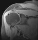
MR of the right shoulder looking at rotator cuff and head of humerus as well as glenoid portion of scapula.
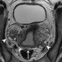
MR image of the pelvis of a woman shows the uterus (arrow) and ovaries (arrowhead).
Pelvic MR image.
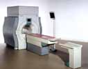
Magnetic Resonance Imaging (MRI) equipment
