Images & Videos: CAT Scan (CT) - Body
Items 11 to 20 of 32 - Click images to view larger
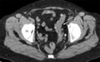
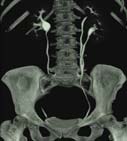
L = Large intestine (colon)
S = Small intestine
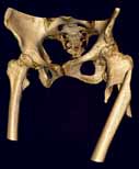
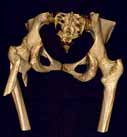
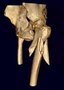
H = Hip joint bones
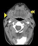
Contrast-enhanced CT of the neck in a patient with lymphoma. Single arrowhead shows abnormal enlarged lymph node in the upper neck on the patient's right side near the angle of the jaw. Double arrowheads show numerous smaller abnormal lymph nodes on the patient's right side.
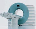
 Patients undergoing a computed tomography (CT) procedure, sometimes called CAT scan, and a newer 64-slice CT scan. The 64-slice CT technology allows the radiologist a greater level of manipulation of 3-D clinical images to provide more complete anatomical views. These visuals and the clinical images can be used to report on CT scans of the body, chest, abdomen, vascular system and head.
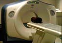
CT urogram demonstrates kidneys, ureters and bladder. This image shows a duplicate ureter on the left. For more information see Urography.
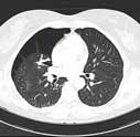
Spontaneous pneumothorax: A cross-section through a patient's chest shows a partially-collapsed lung (arrows). The black space above the edge of the collapsed lung represents air that has leaked from within the lung into the space between the lung and the chest wall.
