Images & Videos: CT Angiography (CTA)
Items 11 to 20 of 53 - Click images to view larger
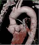
3-D CT angiography image of a patient's aorta. For more information see CT Angiography (CTA).
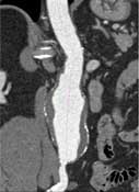
3-D CT angiography image of a patient's aorta. For more information see CT Angiography (CTA).
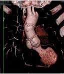
CT angiography showing an enlarged abdominal aorta, also called an aneurysm. For more information see CT Angiography (CTA).
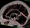
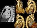
CT angiography images of the brain and arteries to the brain are captured and then reviewed by a radiologist. Angiography can be combined with CT to produce pictures of major blood vessels throughout the body. Angiography uses a contrast material to produce detailed images of both blood vessels and tissues. For more information see CT Angiography (CTA).
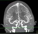
 CT angiography images of the neck and arteries to the brain and face are captured and then reviewed by a radiologist. Angiography can be combined with CT to produce pictures of major blood vessels throughout the body. CT Angiography uses a contrast material to produce detailed images of both blood vessels and tissues.