Images & Videos: Abdominal and Pelvic CT
Items 11 to 20 of 32 - Click images to view larger
 Your Radiologist Explains CT of the Abdomen and Pelvis
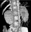
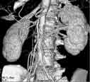
3D-CT of the aorta
3-dimensional reconstruction of a CT scan showing an aortic endograft and femoral artery bypass graft.
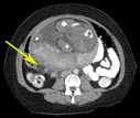
A CT scan of the abdomen and pelvis showing appendicitis (arrow). For more information see Appendicitis.

Computed tomography (CT) scan of an obstructed appendix.
Computed tomography (CT) scan of an obstructed appendix shown lengthwise.
 Your Radiologist Explains Body CT/CAT Scan

Abdominal aorta with atherosclerosis.
3-dimenstional images of the abdominal aorta reconstructed from CT images. Volume rendered images (left) and maximal intensity projection (right) reconstructions show atherosclerotic plaques (arrows) in the origins of the vessels supplying blood to the bowel.
Abdominal aorta and arteries to the kidneys and intestines.

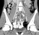
Closed loop small bowel obstruction shown on a CT scan.
