
News from the RSNA Annual Meeting CT Best at Uncovering Drug Mule Payload
December 1, 2010
At A Glance
- CT is the most accurate method of detecting cocaine inside the body of a drug mule.
- Cocaine containers can be as large as a banana or as small as a blueberry.
- Low-dose CT protocols need to be implemented for future routine imaging of suspected drug mules.
- X-rays identified the presence of cocaine containers 70% of the time.
CHICAGO — According to a study presented today at the annual meeting of the Radiological Society of North America (RSNA), the best way to detect cocaine in the body of a human drug courier, known as a mule, is through computed tomography (CT).
"Cocaine from South America is making its way to Europe through Africa," said Patricia Flach, M.D., a radiologist at University Hospital of Berne and Institute of Forensic Medicine of Berne in Switzerland. "From Africa, drug mules most commonly try to enter the European Union and Switzerland."
When legal authorities suspect an individual of being a drug mule, they often turn to radiologists to help quickly detect the presence of cocaine concealed in the body. Cocaine containers, which may be swallowed or inserted in the vagina or rectum, can be as large as a banana or as small as a blueberry.
"In these cases it is important for us to know that we have identified all the drug containers in a body, both for legal purposes and for the health of the patient," Dr. Flach said. "However, there was no research telling us which imaging modality was best in detecting cocaine containers in the stomach, intestines or other body orifices."
Dr. Flach and colleagues analyzed images from 89 exams performed on 50 suspected drug mules over a three-year period at University Hospital. The study group included 45 men and five women between the ages of 16 and 45. Forty-three of the suspects were ultimately identified as drug mules.
Of the imaging exams conducted, 27 were CT, 50 were digital x-ray and 12 were low-dose linear slit digital radiography (LSDR), an extremely fast, high-resolution, full-body x-ray system primarily used for trauma patients. The radiologic findings were compared with a written record of the drug containers recovered from the feces of suspects.
"As we expected, CT imaging allowed us to see all the drug containers, especially when we knew what to look for," Dr. Flach said.
The results showed that the coating and manufacture of the containers changed their appearance, especially on CT images. Rubber coated condoms filled with cocaine appeared very hyper-dense, or white, on CT, while other containers of similar size with plastic foil wrapping appeared iso- to hypo-dense, or gray to black. This contradicts some previous reports that have suggested image density may correlate with the drug content.
The sensitivity of CT was 100 percent, meaning CT was able to find all cocaine containers that were present in the drug mules' bodies. LSDR had a sensitivity rate of 85 percent, and digital x-ray was able to identify the presence of cocaine containers only 70 percent of the time.
"There were positive findings on CT that were clearly not detectable on x-rays due to overlap of intestinal air, feces or other dense structures," Dr. Flach said.
While CT was clearly the most accurate imaging modality in detecting the drug containers, the increased ionizing radiation associated with the exam is a concern when imaging people who are presumably healthy.
"CT is the way to go," Dr. Flach said. "But low-dose protocols need to be implemented to ensure the safety of the people undergoing the procedure."
Coauthors are Steffen Ross, M.D., Gary Hatch, M.D., Ulrich Preiss, M.D., Thomas Ruder, M.D., Michael Thali, M.D., and Michael Patak, M.D.
Video clips (MOV)
- Video clip (4.65 Mbyte)
3_Residual_pack_rectum_body_packer_isodense.mov: Footage showing a reconstruction of a patient's body. A residual pack is visible in the patient's rectum. - Video clip (1.54 Mbyte)
12_1_body_packer_31_slightly_hyperdense-packs_already_excreted_some.mov: Footage showing a 3-D reconstruction of a patient's body. Hyperdense packs are visible in the abdomen. - Video clip (3.44 Mbyte)
8_2_incidental_finding_of_sublingual_cocainepellets.mov: Footage showing a 3-D reconstruction of a patient's head. Sublingual cocaine pellets were incidentally found.
Images (JPG)
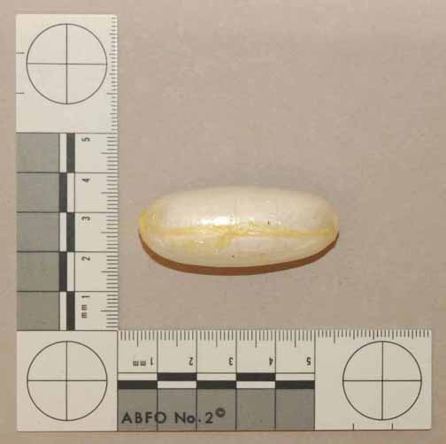
Figure 1: Plastic-wrapped cocaine container (11.5 grams). |
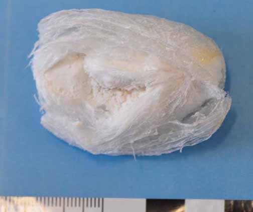
Figure 2: Longitudinally opened package with compressed cocaine paste inside. |

Figure 3: The inner sheath is a thin plastic bag for food knotted and heat-sealed at the point (left). This is wrapped with another plastic bag and sealed the same way. Cellophane is taped, being an intermediate layer (small tape or one broad adhesive foil) and encased in a thicker plastic sheath. The outer layer is again taped plastic foil and the polyethyleyne food wrapping is heat-sealed at one end (middle). Note the neat manufactured drug container (right). |
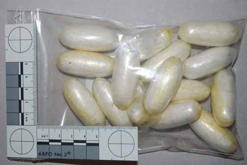
Figure 4: Evidence of cocaine containers found on a drug smuggler. |
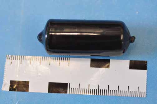
Figure 5: Tightly cocaine-filled condom with bindings on the right end (creating a structure on plain radiographs which is commonly known as the rosette-sign and the typical air filled reservoir on the left side which can be seen on x-ray, too). The condom can be sealed with caoutchouc or rubber. This pack has a weight of 8.8 grams. |
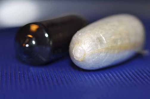
Figure 6: Close-up of a coated condom (black, on the left) and a plastic foil wrapped cocaine container (white, on the right). |
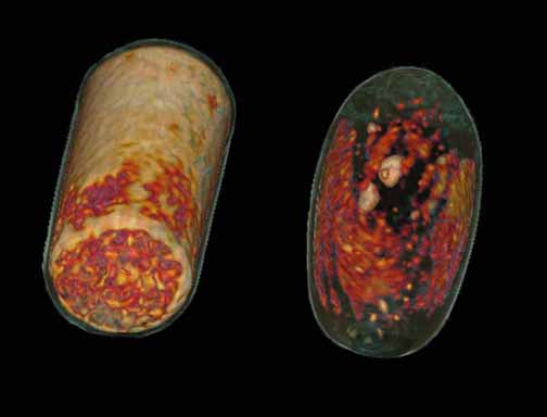
Figure 7: A 3-D reconstruction of the CT dataset, showing the different densities of the same packages as in Figure 6. The rubber-coated condom shows a denser appearance although both packs contain cocaine of high purity. This suggests that measured densities on CT do not imply the content (e.g. methamphetamine, heroin, cocaine or opium) but more the manufacture. |
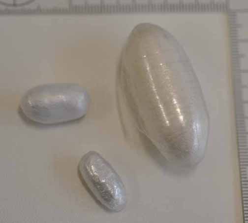
Figure 8: Note the variable appearance of the larger body pack and the smaller pellets usually ingested by users or traffickers in fear of detection by authorities. |

Figure 9: Evidence analyzed at the Forensic Institute of Berne, Switzerland (Virtopsy) after the gastrointestinal passage. Common types of body packs. |
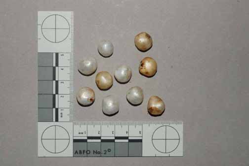
Figure 10: Typical pellets swallowed by body stuffers (approx. 0.5 to 1.0 gram). A tenth of a gram as a single dose is considered lethal in non-users. |
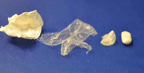
Figure 11: The center core is a small amount of compressed cut or pure cocaine that is covered by a plastic sheath, surrounded by polyethylene food wrapping with a plastic bag around it. Toilet or filter paper is also one of the inner layers. |
Figure 12: A Lodox exam of a body pusher with three packs located in the rectum. |
Figure 13: A Lodox exam of a body pusher; this image shows a magnified view of the pelvis. |
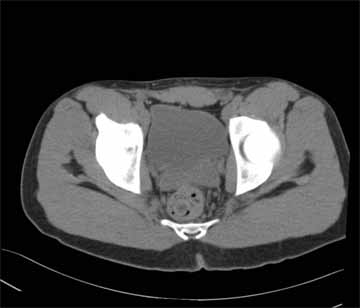
Figure 14: CT scan showing the axial plane at the level of the urine bladder. Follow-up exam after 11 hours by CT revealing one residual isodense cocaine container located in the rectum (the pack had a length of approximately 5 centimeters). This pack was not detectable on prior digital radiograph (DR) due to overlapping structures. |
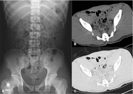
Figure 15: A: A digital radiograph (DR) of the supine view. There are no clearly detectable packs. The diagnosis is aggravated by vast intestinal air and scybala (hardened feces). B: A computed tomography (CT) scan showing the axial plane at the level of the sigmoid colon, abdominal window. C: Same level in lung window. Both (B & C) show a CT performed 17 hours after DR. There were two hypodense packs located in the recto-sigmoid of the unmasked and confessable (after CT) drug mule (only one pack is displayed on this CT slice). |
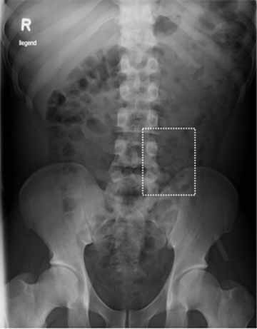
Figure 16: Digital radiograph (DR) of a body stuffer with four hardly detectable pellets paravertebral on the left at the level of the 4th/5th lumbar vertebrae. |
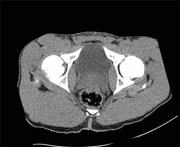
Figure 17: Computed tomography (CT) image of the axial plane, abdominal window. This image was taken of a known drug trafficker. The police witnessed body stuffing of five one-gram cocaine pellets. The image was acquired two days after ingestion. |
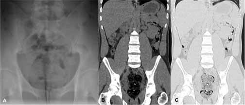
Figure 18: A: Digital radiograph (DR) of the supine view; magnification of the rectal ampoule with obstipated feces and a suspect inhomogeneity projecting on the superior margin of the symphysis. A reliable diagnosis is not feasible; the amount of drug containers is clearly underestimated. B: Computed tomography (CT) scan showing the coronal plane; abdominal window of the rectum. C: CT scan showing the lung window at the same level. Note: one condom filled with several small pellets and two adjacent singular pellets. This finding is hardly assessable in the abdominal window. CT revealed a total of six small pellets and one condom filled with several small cocaine containers. |
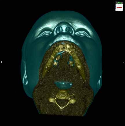
Figure 19: A 3-D reconstruction (view from anterior inferior / view from the chin up to the forehead) of an incidental finding of three sublingual cocaine pellets in a case of brawl/fist fight with subsequent computed tomography (CT) and scan of the viscerocranium. The patient was immediately put under arrest and transferred to the affiliated custody ward. |
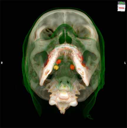
Figure 20: A 3-D reconstruction (view from anterior inferior / view from the chin up to the forehead) of an incidental finding of three sublingual cocaine pellets in a case of brawl/fist fight with subsequent computed tomography (CT) and scan of the viscerocranium. The patient was immediately put under arrest and transferred to the affiliated custody ward. |
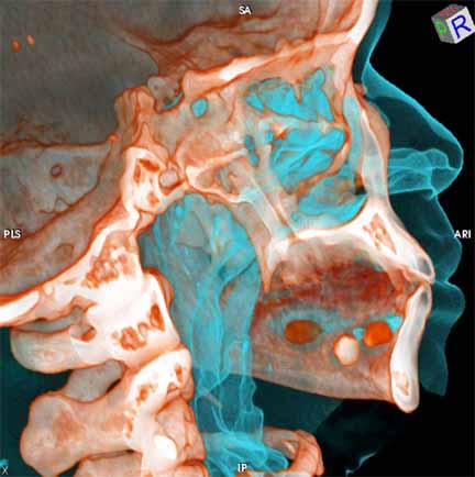
Figure 21: A 3-D reconstruction (sagittal view from the right side) of an incidental finding of three sublingual cocaine pellets in a case of brawl/fist fight with subsequent computed tomography (CT) and scan of the viscerocranium. The patient was immediately put under arrest and transferred to the affiliated custody ward. |
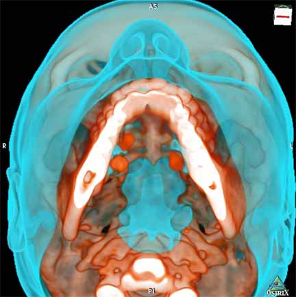
Figure 22: A 3-D reconstruction (view from anterior inferior / view from the chin up to the forehead) of an incidental finding of three sublingual cocaine pellets in a case of brawl/fist fight with subsequent computed tomography (CT) and scan of the viscerocranium. The patient was immediately put under arrest and transferred to the affiliated custody ward. |
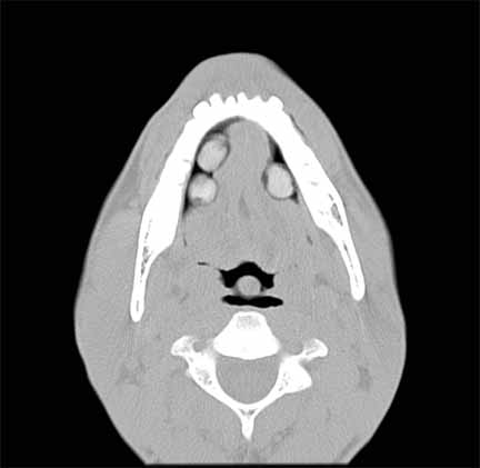
Figure 23: Computed tomography (CT) scan showing the axial plane at the level of the mandible; showing three cocaine pellets underneath the tongue. This scan was performed to rule out facial fractures sustained during a fist fight. |
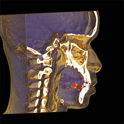
Figure 24: A 3-D reconstruction (sagittal view from the right side) of an incidental finding of three sublingual cocaine pellets in a case of brawl/fist fight with subsequent computed tomography (CT) and scan of the viscerocranium. The patient was immediately put under arrest and transferred to the affiliated custody ward. |
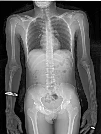
Figure 25: X-ray image of a detained suspect after he swallowed three hidden pellets in his mouth. The patient was then transferred to the affiliated holding cell of the university hospital. Lodox revealed a minimum of six pellets distributed within the stomach and the small intestines. Recovered evidence from the feces showed a total of nine pellets. |
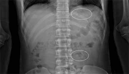
Figure 26 X-ray mage showing a magnified view of the Lodox; pellets are circled. |
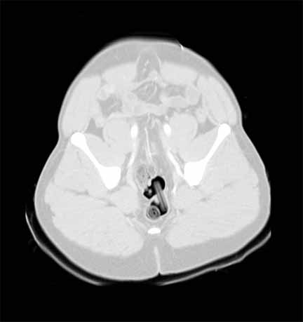
Figure 27: Computed tomography (CT) image of a patient that was brought to the emergency room due to a jump out of a burning house and fall from heights. The patient underwent whole body polytrauma CT protocol. The patient is well known drug traffickers. CT revealed two atypical shaped intra-corporal containers (condoms with unclear content) in the rectum. Recovered evidence showed two condoms filled with a multitude of Swiss Francs money bills, suspected being drug money. This view (oblique plane) shows the pulmonary window. |
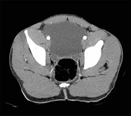
Figure 28: Computed tomography (CT) image of a patient that was brought to the emergency room due to a jump out of a burning house and fall from heights. The patient underwent whole body polytrauma CT protocol. The patient is well known drug traffickers. CT revealed two atypical shaped intra-corporal containers (condoms with unclear content) in the rectum. Recovered evidence showed two condoms filled with a multitude of Swiss Francs money bills, suspected being drug money. In this view (oblique plane), the money container is not detectable. |
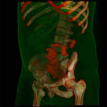
Figure 29: A 3-D reconstruction of the body. This image illustrates a body packer with a multitude of residual packs mainly located in the recto-sigmoid bowel. |
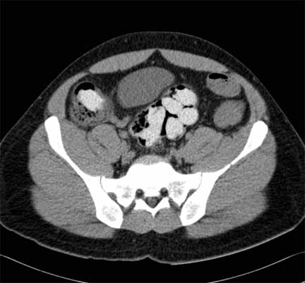
Figure 30: Computed tomography (CT) image depicting slightly hyperdense cocaine containers within the alimentary tract. |