Obstetric Ultrasound

- What is Obstetrical Ultrasound Imaging?
- What are some common uses of the procedure?
- How should I prepare?
- What does the equipment look like?
- How does the procedure work?
- How is the procedure performed?
- What will I experience during and after the procedure?
- Who interprets the results and how do I get them?
- What are the benefits vs. risks?
- What are the limitations of Obstetrical Ultrasound Imaging?
What is Obstetrical Ultrasound Imaging?
Ultrasound is safe and painless, and produces pictures of the inside of the body using sound waves. Ultrasound imaging, also called ultrasound scanning or sonography, involves the use of a small transducer (probe) and ultrasound gel placed directly on the skin. High-frequency sound waves are transmitted from the probe through the gel into the body. The transducer collects the sounds that bounce back and a computer then uses those sound waves to create an image. Ultrasound examinations do not use ionizing radiation (as used in x-rays), thus there is no radiation exposure to the patient. Because ultrasound images are captured in real-time, they can show the structure and movement of the body's internal organs, as well as blood flowing through blood vessels.
Ultrasound imaging is a noninvasive medical test that helps physicians diagnose and treat medical conditions.
Obstetrical ultrasound provides pictures of an embryo or fetus within a woman's uterus, as well as the mother's uterus and ovaries.
A Doppler ultrasound study may be part of an obstetrical ultrasound examination.
Doppler ultrasound is a special ultrasound technique that evaluates blood flow through a blood vessel, including the body's major arteries and veins in the abdomen, arms, legs, neck and head (in infants and children).
During an obstetrical ultrasound the examiner may evaluate blood flow in the umbilical cord or may, in some cases, assess blood flow in the fetus or placenta.
What are some common uses of the procedure?


Obstetrical ultrasound is a useful clinical test to:
- establish the presence of a living embryo/fetus
- estimate the age of the pregnancy
- diagnose congenital abnormalities of the fetus
- evaluate the position of the fetus
- evaluate the position of the placenta
- determine if there are multiple pregnancies
- determine the amount of amniotic fluid around the baby
- check for opening or shortening of the cervix
- assess fetal growth
- assess fetal well-being
Some physicians also use 3-D ultrasound to image the fetus and determine if it is developing normally.
How should I prepare?
You should wear a loose-fitting, two-piece outfit for the examination. Only the lower abdominal area needs to be exposed during this procedure.
The radiologist or sonographer may elect to examine an early pregnancy by means of transvaginal ultrasound in order to see the pregnancy more closely or to assess the cervix. For more information on transvaginal ultrasound, see the Pelvic Ultrasound page (www.RadiologyInfo.org/en/info.cfm?pg=pelvus).
What does the equipment look like?

Ultrasound scanners consist of a console containing a computer and electronics, a video display screen and a transducer that is used to do the scanning. The transducer is a small hand-held device that resembles a microphone, attached to the scanner by a cord. The transducer sends out inaudible high frequency sound waves into the body and then listens for the returning echoes from the tissues in the body. The principles are similar to sonar used by boats and submarines.
The ultrasound image is immediately visible on a video display screen that looks like a computer or television monitor. The image is created based on the amplitude (loudness), frequency (pitch) and time it takes for the ultrasound signal to return from the area of the patient being examined to the transducer (the device used to examine the patient), as well as the type of body structure and composition of body tissue through which the sound travels. A small amount of gel is put on the skin to allow the sound waves to travel back and forth from the transducer.
How does the procedure work?
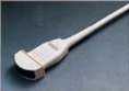
Transabdominal transducer
Ultrasound imaging is based on the same principles involved in the sonar used by bats, ships and fishermen. When a sound wave strikes an object, it bounces back, or echoes. By measuring these echo waves, it is possible to determine how far away the object is as well as the object's size, shape and consistency (whether the object is solid or filled with fluid).
In medicine, ultrasound is used to detect changes in appearance, size or contour of organs, tissues, and vessels or detect abnormal masses, such as tumors.
In an ultrasound examination, a transducer both sends the sound waves and receives the echoing waves. When the transducer is pressed against the skin, it directs small pulses of inaudible, high-frequency sound waves into the body. As the sound waves bounce off internal organs, fluids and tissues, the sensitive microphone in the transducer records tiny changes in the sound's pitch and direction. These signature waves are instantly measured and displayed by a computer, which in turn creates a real-time picture on the monitor. One or more frames of the moving pictures are typically captured as still images. Small loops of the moving “real time” images may also be saved.

The movement of the embryo or fetus and his or her heartbeat can be seen as an ongoing ultrasound movie. Most ultrasound devices also have an audio component that processes the echoes produced by blood flowing through the fetal heart, blood vessels and umbilical cord. This sound can be made audible to human ears and has been described by patients as a whooshing noise.
Doppler ultrasound, a special application of ultrasound, measures the direction and speed of blood cells as they move through vessels. The movement of blood cells causes a change in pitch of the reflected sound waves (called the Doppler effect). A computer collects and processes the sounds and creates graphs or color pictures that represent the flow of blood through the blood vessels.
How is the procedure performed?

For most ultrasound exams, you will be positioned lying face-up on an examination table that can be tilted or moved.
After you are positioned on the examination table, the radiologist or sonographer will apply a warm water-based gel to the area of the body being studied. The gel will help the transducer make secure contact with the body and eliminate air pockets between the transducer and the skin that can block the sound waves from passing into your body. The transducer is placed on the body and moved back and forth over the area of interest until the desired images are captured.
There is usually no discomfort from pressure as the transducer is pressed against the area being examined. However, if scanning is performed over an area of tenderness, you may feel pressure or minor pain from the transducer.
Once the imaging is complete, the clear ultrasound gel will be wiped off your skin. Any portions that are not wiped off will dry to a powder. The ultrasound gel does not stain or discolor clothing.
Sometimes the radiologist determines that a transvaginal scan needs to be performed. This technique often provides improved, more detailed images of the uterus and ovaries. This method of scanning is especially useful in early pregnancy.
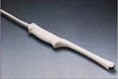
Transvaginal transducer
Transvaginal ultrasound is performed very much like a gynecologic exam and involves the insertion of the transducer into the vagina after you empty your bladder. The tip of the transducer is smaller than the standard speculum used when performing a Pap test. A protective cover is placed over the transducer, lubricated with a small amount of gel, and then inserted into the vagina. Only two to three inches of the transducer end are inserted into the vagina. The images are obtained from different orientations to get the best views of the uterus and ovaries. Transvaginal ultrasound is usually performed with you lying on your back, possibly with your feet in stirrups similar to a gynecologic exam.
Doppler sonography is performed using the same transducer.
What will I experience during and after the procedure?

Ultrasound examinations are painless and easily tolerated by most patients.
However, at times during an obstetrical ultrasound, the sonographer may have to press more firmly to get closer to the embryo or fetus to visualize the structure better. Any discomfort is usually minimal and temporary.
At times the sonographer may have to press more firmly to get closer to the embryo or fetus to visualize the structure better. Any discomfort is usually minimal and temporary.
If a Doppler ultrasound study is performed, you may actually hear pulse-like sounds that change in pitch as the blood flow is monitored and measured.
With transvaginal scanning, there may be minimal discomfort as the transducer is inserted into the vagina.
This ultrasound examination is usually completed within 30 minutes.
When the examination is complete, you may be asked to dress and wait while the ultrasound images are reviewed.
After an ultrasound examination, you should be able to resume your normal activities immediately.
Who interprets the results and how do I get them?
A radiologist, a physician specifically trained to supervise and interpret radiology examinations, will analyze the images and send a signed report to your primary care physician, or to the physician or other healthcare provider who requested the exam, and he/she will share the results with you. In some cases the radiologist may discuss results with you at the conclusion of your examination.
Follow-up examinations may be necessary, and your doctor will explain the exact reason why another exam is requested. Sometimes a follow-up exam is done because a suspicious or questionable finding needs clarification with additional views or a special imaging technique. A follow-up examination may also be necessary so that any change in a known abnormality can be monitored over time. Follow-up examinations are sometimes the best way to see if treatment is working or if an abnormality is stable over time.
What are the benefits vs. risks?
Benefits
- Most ultrasound scanning is noninvasive (no needles or injections).
- Occasionally, an ultrasound exam may be temporarily uncomfortable, but it is almost never painful.
- Ultrasound is widely available, easy-to-use and less expensive than other imaging methods.
- Ultrasound imaging is extremely safe and does not use any ionizing radiation.
- Ultrasound scanning gives a clear picture of soft tissues that do not show up well on x-ray images.
- Ultrasound is the preferred imaging modality for the diagnosis and monitoring of pregnant women and their unborn babies.
- Ultrasound has been used to evaluate pregnancy for nearly four decades and there has been no evidence of harm to the patient, embryo or fetus. Nevertheless, ultrasound should be performed only when medically indicated.
- Ultrasound allows the doctor to see inside the uterus and provides much information about the pregnancy.
Risks
- For standard diagnostic ultrasound, there are no known harmful effects on humans.
What are the limitations of Obstetrical Ultrasound Imaging?
Obstetric ultrasound cannot identify all fetal abnormalities. Consequently, when there are clinical or laboratory suspicions for a possible abnormality, a pregnant woman may have to undergo nonradiologic testing such as amniocentesis (the evaluation of fluid taken from the sac surrounding the fetus) or chorionic villus sampling (evaluation of placental tissue) to determine the health of the fetus, or she may be referred by her primary care provider to a perinatologist (an obstetrician specializing in high-risk pregnancies).
Locate an ACR-accredited provider: To locate a medical imaging or radiation oncology provider in your community, you can search the ACR-accredited facilities database.
This website does not provide costs for exams. The costs for specific medical imaging tests and treatments vary widely across geographic regions. Many—but not all—imaging procedures are covered by insurance. Discuss the fees associated with your medical imaging procedure with your doctor and/or the medical facility staff to get a better understanding of the portions covered by insurance and the possible charges that you will incur.
Web page review process: This Web page is reviewed regularly by a physician with expertise in the medical area presented and is further reviewed by committees from the American College of Radiology (ACR) and the Radiological Society of North America (RSNA), comprising physicians with expertise in several radiologic areas.
Outside links: For the convenience of our users, RadiologyInfo.org provides links to relevant websites. RadiologyInfo.org, ACR and RSNA are not responsible for the content contained on the web pages found at these links.
Images: Images are shown for illustrative purposes. Do not attempt to draw conclusions or make diagnoses by comparing these images to other medical images, particularly your own. Only qualified physicians should interpret images; the radiologist is the physician expert trained in medical imaging.
|
|
Images/videos |
|
This page was reviewed on July 16, 2013
