November 27, 2007
Forensics Go High-Tech with CT Autopsies
At A Glance
- A new study examines the use of CT in civilian medical examiner autopsies in the U.S.
- CT is a sensitive imaging tool for detecting injuries and the cause of death in victims of blunt trauma.
- CT autopsy is noninvasive and saves time and money compared to conventional autopsy.
CHICAGO — Radiologists are investigating the use of computed tomography (CT) as a tool for civilian medical examiners' autopsies in the United States. According to findings presented today at the annual meeting of the Radiological Society of North America (RSNA), CT autopsy has the potential to replace conventional autopsy in determining the cause of certain accidental deaths.
"CT is a sensitive imaging tool for detecting injuries and cause of death in victims of blunt trauma," said Barry Daly, M.D., professor of radiology at the University of Maryland School of Medicine and radiologist at the University of Maryland Medical Center in Baltimore. "When there are major injuries, such as those resulting from a motor vehicle accident, CT may provide enough information to enable a conventional autopsy to be avoided altogether."
All states are required by law to perform an autopsy in cases of sudden and unexplained deaths. Of the 8,000 such deaths referred to the chief medical examiner of the state of Maryland last year, approximately one-half required full autopsy.
CT autopsy compares favorably to conventional autopsy in several ways. In cases of suspicious death, the noninvasive procedure does not damage or destroy key forensic evidence, as can happen during a conventional autopsy. In addition, CT can be used in situations where autopsy may be prohibited by religious or cultural beliefs. CT autopsy is considerably less expensive than conventional autopsy and can be performed in a fraction of the time. A forensic medical examiner requires several hours to conduct a full autopsy, while multi-detector CT scanning and interpretation can be completed in about 30 minutes.
In Dr. Daly's study, 20 autopsies were performed using whole-body multi-detector CT. Interpretations of the CT scans by two radiologists were compared with the results of a conventional autopsy performed on each body by state forensic medical examiners. Included were 14 victims of blunt trauma and six victims of a penetrating wound made either by a knife or ballistic weapon.
In all 14 blunt trauma cases and five of the six penetrating wounds, CT accurately identified the cause of death. The radiologists and forensic medical examiners evaluated the CT findings as comparable to conventional autopsy in 13 of the 14 blunt trauma cases and as a helpful adjunct in five of the six penetrating wound cases. In the study, CT was able to localize rapidly all 26 major ballistic fragments recovered from the victims during conventional autopsy.
"Autopsy is mandatory in deaths involving gunshot wounds, but CT can serve as a powerful adjunct to the conventional exam," Dr. Daly said. "Performing CT imaging first may speed up a conventional autopsy, especially when it comes to locating ballistic fragments, which are so important to criminal investigations."
In addition, CT was more sensitive than conventional autopsy in identifying air embolism, an often undetected important contributing factor in fatal trauma.
While CT has previously been used in autopsies of American soldiers and in a few countries outside the U.S., the technology is only now generating strong interest within the nation's forensic community.
"Although these preliminary results are promising, more research is needed to show that CT could be widely used within the U.S. medical examiners system," Dr. Daly said.
Video clips:
- Video clip (AVI format, 4,850 Kbyte)
Multiple skull fractures are demonstrated in a decedent following major trauma sustained in a motor vehicle accident.
Images:
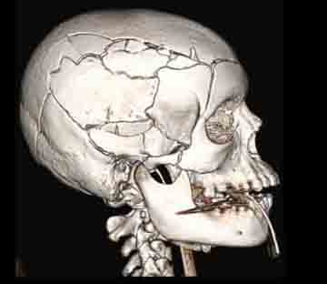 Figure 1. CT image shows multiple skull and facial bone fractures resulting from fatal blunt trauma to the head in a pedestrian struck by a car. |
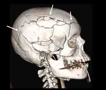 Figure 2. CT image shows multiple skull and facial bone fractures resulting from fatal blunt trauma to the head in a pedestrian struck by a car. Arrows point to some of the fractures. |
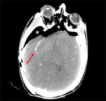 Figure 3. CT image depicting internal damage to the brain with diffuse cerebral injury. Blood is present in the right sylvian fissure (arrow). Air (*) has entered the skull due to the skull fractures. |
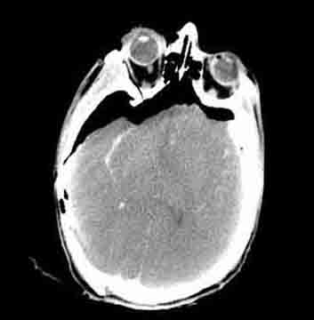 Figure 4. CT image depicting internal damage to the brain with diffuse cerebral injury. Blood is present in the right sylvian fissure. Air has entered the skull due to the skull fractures. |
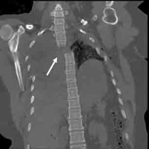 Figure 5. CT image shows a severe displaced fracture through the third upper thoracic spine (arrow) in a decedent who suffered multiple injuries in a motor vehicle accident. |
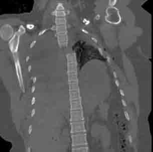 Figure 6. CT image shows a severe displaced fracture through the third upper thoracic spine in a decedent who suffered multiple injuries in a motor vehicle accident. |
 Figure 7. Image of CT machinery |
# # #
